
Fig. 6
- ID
- ZDB-FIG-131220-32
- Publication
- Yasuda et al., 2013 - A cis-acting element in the coding region of cyclin B1 mRNA couples subcellular localization to translational timing
- Other Figures
- All Figure Page
- Back to All Figure Page
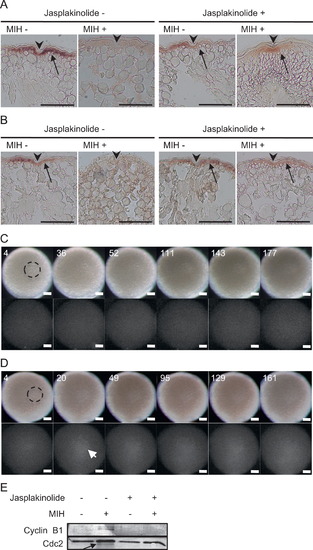
Effects of actin filament stabilization on cyclin B1 mRNA aggregation and translational regulation. ((A), (B)) Section in situ hybridization of wild-type oocytes probed with cyclin B1 (A) and tgo3′ mRNA-expressing oocytes probed with gfp (B). Oocytes were treated with jasplakinolide (+) or DMSO () and stimulated with (+) or without () MIH. The oocytes were fixed 140 min after MIH stimulation. Arrowheads indicate the micro-pile. Arrows indicate the signals of cyclin B1 mRNA (A) and tgo3′ reporter mRNA (B). ((C), (D)) Real-time imaging of oocytes expressing tgo3′ (C) or tgoM3′ mRNAs (D) treated with jasplakinolide and MIH. The times after MIH treatment (min) are shown. Bars, 100 μm. Dotted circles show the GV. Arrow indicates the initial translation signal. Similar results were obtained from six oocytes expressing tgo3′ mRNAs and three oocytes expressing tgoM3′ mRNAs. (E) Anti-Cyclin B1 and anti-Cdc2 immunoblots of oocytes treated with jasplakinolide (+) or DMSO (-) in the presence (+) or absence () of MIH. Cdc2 protein is a loading control of this experiment. An arrow indicates an active form of Cdc2 (Kondo et al., 2001). |
Reprinted from Developmental Biology, 382(2), Yasuda, K., Kotani, T., and Yamashita, M., A cis-acting element in the coding region of cyclin B1 mRNA couples subcellular localization to translational timing, 517-29, Copyright (2013) with permission from Elsevier. Full text @ Dev. Biol.