|
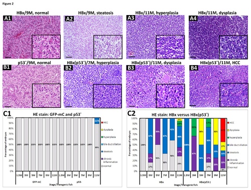
(A1~A4) H&E staining of the liver sections from HBx transgenic fish revealed various pathological phenotypes, such as normal tissue, steatosis, hyperplasia and dysplasia. (B1) H&E staining of the liver sections from p53 mutant fish appeared normal. (B2~B4) H&E staining from the liver sections of HBx(p53-) transgenic fish revealed hyperplasia, dysplasia and HCC. All specimens are shown under 200-fold magnification. Scale bars: 50μm. The boxed areas are enlarged images with 400-fold magnification and are shown in the lower right corner. (C1) Statistical analysis of H&E staining results from GFP-mC and p53 mutant control fish. (C2) Statistical analysis of H&E staining results from HBx and HBx(p53-) transgenic fish. The different colors denote the different pathological features, as follows: gray-normal, purple-chronic inflammation, blue-steatosis, light blue-bile duct dilation, green-hyperplasia, yellow-dysplasia, and red-HCC, respectively.
|