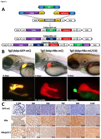
(A) A schematic diagram of the LR recombination reaction used to generate the expression constructs, including three entry clones (p5E-l-fabp, pME-GFP and p3E-mCherry) and two destination vectors (pDestTol2pA and pDestTol2CG2) that contain the cmlc2:GFP-pA expression cassette. The three final constructs are shown at the bottom of the figure. (B) The expression of the GFP-mCherry fusion protein and the HBx-mCherry fusion protein in the liver are shown in four-day-old embryos and eleven-month-old adult wild-type fish harboring the l-fabp:GFP-mCherry transgene and the l-fabp:HBx-mCherry transgene, respectively. The HBx-mCherry fusion protein is expressed in the liver of p53 mutant fish harboring the l-fabp:HBx-mCherry;cmcl2:GFP transgene, as indicated by the red fluorescence, and cmlc2:GFP is expressed in the heart, as indicated by the green fluorescence. (C) HBx protein expression in the hepatocytes was detected via immunostaining of the liver sections of the 1.5-, 3-, 5-, 7-, 9- and 11-month-old fish harboring the l-fabp:HBx-mCherry transgene in the wild-type background or the l-fabp:HBx-mCherry;cmcl2:GFP transgene in the p53 mutant background (x 400). Scale bars: 50 μm.
|
