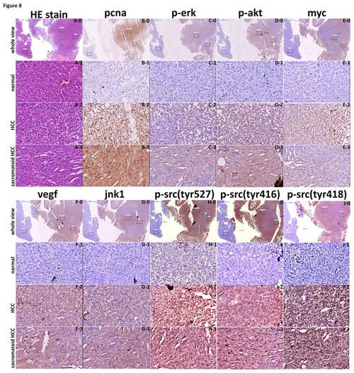
Assessment of pcna, p-erk, p-akt, myc, vegf, jnk1, p-src(tyr527), p-src(tyr416), and p-src(tyr418) signaling in HCC, sarcomatoid HCC, and adjacent normal tissue of src-overexpressing transgenic zebrafish. Liver sections from the src-overexpressing transgenic zebrafish that had developed HCC and sarcomatoid HCC, as well as adjacent normal tissue, were analyzed with H&E staining (A) and immunostaining for pcna, p-erk, p-akt, myc, vegf, jnk1, p-src(tyr527), p-src(tyr416), and p-src(tyr418) (B~J). All slides, comprising normal tissue (1), HCC (2) and sarcomatoid HCC (3), were analyzed using Panoramic Viewer under lower resolution (0) and subsequently at higher resolution. (A0-A3) H&E stain, (B0~B3) pcna protein expression, (C0~C3) p-erk expression level, (D0~D3) p-akt expression level, (E0~E3) myc protein expression, (F0~F3) vegf protein expression, (G0~G3) jnk1 protein expression, (HG0~H3) p-src(tyr527) level, (I0~I3) p-src(tyr416) level, and (J0~J3) p-src(tyr418) expression level were assessed. Scale bars: 50 μm.
|
