
Fig. S1
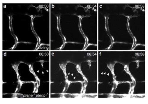
Endothelial cells lacking Pten display protruding filopodia from 75 hpf onwards. Still frames from time lapse movies of Tg(kdrl:eGFP) sibling (Suppl Mov 1, panel a-c) and ptena-/-ptenb-/- mutant embryo (Suppl Mov 2, panel d-f) showing two intersegmental vessels in the trunk of the embryo. Endothelial cells in ptena-/-ptenb-/- mutants display filopodia formation (arrowheads), whereas endothelial cells in siblings do not. Images were taken every 2 minutes, maximum projections of z-stacks (1 μm step size) were used to generate the time lapse movies. Anterior to the left, 40x with 2 zoom, time is indicated in hours and minutes. |