
Fig. S1
- ID
- ZDB-FIG-130826-43
- Publication
- Huang et al., 2013 - Igf Signaling is Required for Cardiomyocyte Proliferation during Zebrafish Heart Development and Regeneration
- Other Figures
- All Figure Page
- Back to All Figure Page
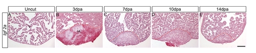
igf2a expression is not detected during zebrafish heart regeneration. ISH was performed on uncut hearts (A) and 3 dpa (B), 7 dpa (C), 10 dpa (D) and 14 dpa (E) regenerating hearts. There is no detectable igf2a expression in regenerating hearts by ISH. Scale bar = 100 μm. v: ventricle; ia: injured area. It was shown previously that embryos injected with igf1r morpholinos exhibited severely reduced body length at 24 hpf [43], raising the possibility that blocking Igf signaling inhibits overall embryonic growth and development in general. We examined embryo length and overall development of Tg(hsp70:dnigf1ra-GFP) embryos or embryos treated with the Igf1r chemical inhibitor NVP-AEW-541 from 48–72 hpf. We observed only a very mild reduction in the length of the inhibitor treated (3.7%) and transgenic embryos (5.7%) (Figure S2. A-E). These results suggest that the reduced cardiomyocyte numbers are unlikely caused by effects of Igf signaling on overall embryo growth and development indicating Igf signaling is required for zebrafish heart development. |