
Fig. 7
- ID
- ZDB-FIG-130826-41
- Publication
- Huang et al., 2013 - Igf Signaling is Required for Cardiomyocyte Proliferation during Zebrafish Heart Development and Regeneration
- Other Figures
- All Figure Page
- Back to All Figure Page
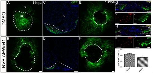
Igf signaling is required for contribution ofgata4:EGFP positive cardiomyocytes to the regenerating area. gata4:EGFP fish were treated with the Igf1r inhibitor NVP-AEW541 from 2-14 dpa (n = 12) (B, D) and 7-10 dpa (n = 5) (F, H). DMSO was used as a control (14dpa: n = 10; 10 dpa: n = 6) (A, C, E, G). Whole mount confocal microscopy from the view of the apex (A, B, E, F) and frozen sections (C, D, G, H) were performed at 14 and 10 dpa to determine the contribution of the EGFP positive population. BrdU (red) and Gata4 (green) double positive cells indicate proliferating gata4:EGFP positive cardiomyocytes (G, G′, H, H′). G′, and H′ are the higher magnification images of the dashed boxes in G and H. BrdU staining (red) and gata4:EGFP (green) were shown as separated channel images. The dashed line marks the regenerating area. Scale bar: (B, D, F, H) = 50 μm; (H′) = 20 μm. ia: injured area, v: ventricle. (I) Quantification of BrdU and gata4:EGFP double positive cells/gata4:EGFP area ± S.E. A significant decrease (*p<0.05) in gata4:EGFP positive cell proliferation was detected in fish treated with Igf1r inhibitor NVP-AEW541 from 7-10dpa. |