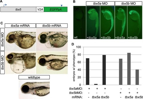
mRNA injection rescue studies indicate that tbx5a and tbx5b are not functionally redundant in cardiac development. A: Schematic of mRNA structure for rescue constructs, in which the coding sequence for tbx5a or tbx5b was connected to enhanced green fluorescent protein (EGFP, with accompanying poly-adenylation signal) by means of a viral 2A peptide. Blue arrow indicates location of translation start site. Blue arrowhead indicates termination signal. B: Fluorescent images of 26 hours postfertilization (hpf) embryos previously injected with combinations of either buffer (in wild-type, wt), tbx5aMO or tbx5bMO, and tbx5a or tbx5b mRNA to evaluate mRNA integrity by means of production of translated GFP. C: Brightfield images of 72 hpf embryos showing representative cardiac phenotypes for each group of injected embryos. D: Quantification of morpholino injected embryos displaying a cardiac morphology phenotype at 54 hpf. (tbx5aMO 70.3% n = 91; tbx5a MO+ tbx5a mRNA 2.4% n = 84; tbx5a MO+ tbx5b mRNA 78.9% n = 133; tbx5b MO 69.1% n = 220; tbx5b MO+ tbx5a mRNA 84.6% n = 65, tbx5b MO+ tbx5b mRNA 40.6% n = 69).
|
