
Fig. 7
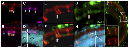
Regeneration of UV cones following prodrug cell ablation. Regenerating UV cones were observed in larvae 5 days after MTZ treatment. Co-localization of these UV cone markers with BrdU detection indicated that the UV cones were recently proliferating. The right-most cell (A–C) is double labelled for BrdU and UV opsin (mCherry) (filled arrow with red outline; has not yet differentiated to a cone morphology). Other UV cones have begun to reappear (empty red arrows), presumably outside of the window of BrdU application. Other BrdU-positive photoreceptors are detectable (filled arrows), likely representing rods, nascent UV cones, or UV cones not expressing the transgene. One week after MTZ ablation, the regeneration of morphologically mature cones was observed (E–H). BrdU+ cells co-labelled with UV cone mCherry expression (E–G; arrow). The regenerated UV cones are morphologically similar to newly generated UV cones in the expanding retinal margin (I), demonstrating that the regenerated cones are qualitatively normal. Scale bars for A–H = 5 µm, J = 50 µm. |
| Gene: | |
|---|---|
| Fish: | |
| Condition: | |
| Anatomical Term: | |
| Stage Range: | Days 7-13 to Days 14-20 |