|
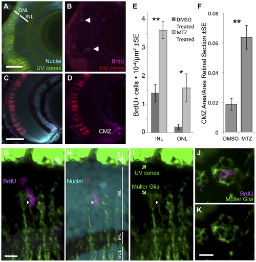
Ablation of cones, primarily UV cones, is Our transgenic UV cone ablation model fish treated with prodrug MTZ showed a significant increase in proliferating cells in the retina at 24 hours after ablation (A, B, especially notice cells demarcated with arrowheads in top right away, from the retinal margin (ciliary marginal zone, CMZ)) compared to sibling fish treated with a DMSO vehicle control solution (C, D). BrdU is incorporated into the CMZ of all fish with and without cone ablation, as expected (A–D). Proliferating cells were quantified in the inner and outer nuclear layers (INL and ONL) at 24 hours after ablation (E) (**INL p = 0.004, *ONL p = 0.002, DMSO treated n = 9, MTZ treated n = 10). The area of the CMZ was also greater in extent after MTZ treatment, calculated relative to the area of the entire retinal sections (F, **p<0.001, MTZ-treated n = 11, DMSO treated n = 15). Scale bars = 5 & 3 µm in A & C, respectively. G–I. The proliferating cells that increase in abundance during regeneration include Müller glia in the INL, as indicated by close apposition of Müller glia markers (green) with BrdU+ nuclei (magenta) (arrowhead). Example shown is from Movie S1 with a cocktail of two antibodies against Müller glia (green, anti-GFAP & anti-glutamine synthetase, both raised in mouse. Saturated green at top of figure is from UV cones expressing abundant GFP, scale bar = 5 µm) and anti-glutamine synthetase antibody produced similar results alone (Movie S2). ONL, outer nuclear layer; INL, inner nuclear layer; IPL, inner plexiform layer; GCL, ganglion cell layer. J–K. Same labelling regimes as panel G but as a tangential section (orthogonal to G and equivalent plane to Figure 1), showing BrdU+ nuclei in the INL surrounded by Müller glia. Scale bar = 5 µm.
|