
Fig. 1
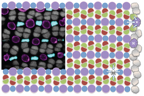
Zebrafish cone photoreceptor mosaic and experimental rationale. The cone photoreceptor mosaic of zebrafish consists of a precise reiterated arrangement of cone spectral sensitivity subtypes, extending in rows radiating toward the periphery of the retina (towards the right). The inset micrograph demonstrates the positions of the UV- and blue-sensitive cones (pseudocoloured magenta and blue, respectively, in a transgenic fish with these cone subtypes expressing GFP and mCherry), and the nuclei of green- and red-sensitive cones forming rows in between (grey). This tangential view of the cones is orthogonal compared to Figure 2, at a level just above the magenta BrdU+ nuclei in panel 2E. This mosaic is schematized, with magenta circles representing the UV cones and other coloured shapes representing their respective cone sensitivities. New cones are continuously added to the periphery of the adult retina from stem cells near the iris (cloud shapes on right); thus the existing mosaic serves as a template for specifying cone identity and/or position of newly added retina. Towards the top right one can imagine a UV cone (*) during differentiation, and its specification being influenced by the identity of the existing mosaic (arrows). Alternatively, mathematical modelling also supports that newly generated cones could be specified without regard for position (e.g. differentiating UV cone marked with adjacent to an existing UV cone, this arrangement is not observed in mature mosaic) and subsequently move to their correct position. Our intervention principally ablates UV cones (skull & cross-bones, ) and hypothesizes that the fate of the regenerating cell will be influenced (arrows) by the remaining cones. |