
Fig. 1
- ID
- ZDB-FIG-130218-25
- Publication
- Janssens et al., 2013 - Matrix metalloproteinase 14 in the zebrafish: an eye on retinal and retinotectal development
- Other Figures
- All Figure Page
- Back to All Figure Page
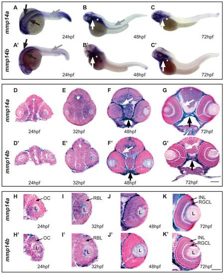
Spatiotemporal expression pattern of mmp14a and mmp14b mRNA in developing zebrafish. A-C & A′-C′ Whole mount in situ hybridization (ISH) for mmp14a (A-C) and mmp14b (A′-C′) in zebrafish embryos at various developmental stages shows mmp14a and mmp14b expression in the head mesenchyme lateral and ventro-lateral to the hindbrain (black arrows in A and A2) at 24 hpf, as well as in myosepta in the trunk and tail (grey arrow in A, B and A2) at 24 hpf and 48 hpf. Mmp14a and mmp14b expression in craniofacial elements (white arrow in B-C, B′-C′) and pectoral fins (white star in B-C, B′-C′) is apparent from 48 hpf onwards. D-G & D′-G′ Transverse sections through the head of zebrafish embryos, stained via whole mount ISH for mmp14a (D-G) and mmp14b (D2-G2) and counterstained with Nuclear Fast Red, show mRNA expression in the brain, cartilage (black arrow) and connective tissue (white arrow) at various developmental stages. H-K & H′-K′ Detailed view of the retina at various developmental stages shows prominent mmp14a expression in the optic cup (H) at 24 hpf, the retinoblast layer (I, J) at 32 and 48 hpf and in the INL and RGCL (K) at 72 hpf, while mmp14b expression in the retina is almost absent (H′-K′). hpf, hours post fertilization; INL, inner nuclear layer; L, lens; OC, optic cup; RBL, retinoblast layer; RGCL, retinal ganglion cell layer. Scale bars: 50 μm. |
| Genes: | |
|---|---|
| Fish: | |
| Anatomical Terms: | |
| Stage Range: | Prim-5 to Protruding-mouth |