
Fig. 6
- ID
- ZDB-FIG-130206-1
- Publication
- Li et al., 2012 - Reciprocal Regulation between Resting Microglial Dynamics and Neuronal Activity In Vivo
- Other Figures
- All Figure Page
- Back to All Figure Page
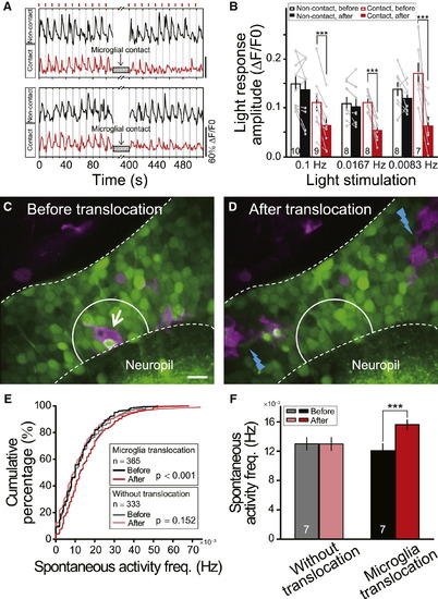
Microglial Contact Reduces Visually Evoked Responses of Tectal Neurons(A) Examples of repeated moving bar-evoked Ca2+ activities of tectal neurons contacted (Contact, red) or not contacted (Non-contact, black) by microglial bulbous endings. Short red bars (top) indicate the moving bar stimuli applied at 0.1 Hz.(B) Summary of light responses in tectal neurons evoked by moving bars with frequencies of 0.1 Hz, 0.0167 Hz, and 0.0083 Hz. Data after microglial contacts (red filled bars) in three sets of experiments were obtained during 0–100, 0–360, and 0–480 s after the contact, respectively. Numbers of tectal neurons examined on bar. Each microglia-contacted neuron was from an individual larva, and at least one neuron without microglial contacts was examined simultaneously.(C and D) Position of one microglial cell (arrow in C) before and after two-photon laser-induced local injury in nearby regions (blue lightning in D). Images from one optical section in optic tectum of Tg(Coronin1a:DsRed) larva loaded with OGB-AM. Area between white dashed lines, optic tectum soma layer. Scale bar represents 10 μm.(E) Cumulative distribution of spontaneous Ca2+ activity frequency of neurons in truncated white circles (20 μm diameter) in (C) and (D) before and after laser-induced injury, which led to translocation (7 out of 14 larvae) or no translocation (7 out of 14 larvae) of microglia examined. Numbers of neurons examined in boxes.(F) Summary data showing spontaneous Ca2+ activity frequency in microglial pre-existing area before and after microglial translocation. Numbers of larvae examined on bars. p < 0.001. Error bars represent ± SEM.See also Figure S5. |
Reprinted from Developmental Cell, 23(6), Li, Y., Du, X.F., Liu, C.S., Wen, Z.L., and Du, J.L., Reciprocal Regulation between Resting Microglial Dynamics and Neuronal Activity In Vivo, 1189-1202, Copyright (2012) with permission from Elsevier. Full text @ Dev. Cell