
Fig. 8
- ID
- ZDB-FIG-130124-7
- Publication
- Vick et al., 2012 - Learning the Scientific Method Using GloFish
- Other Figures
- All Figure Page
- Back to All Figure Page
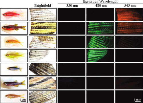
Different colored GloFish have distinct patterns of fluorescence in their caudal fins. GloFish with different body colors under regular room light were assayed for fluorescence emission after excitation by different wavelengths of light. Each row contains images of the same fish. The first column has images of the whole fish, and the remaining columns have images of the surgically removed caudal fin. A range of wavelengths was used to excite the fluorophores, and the peak wavelength is listed above each column. The fins are in lateral views with anterior to the left and dorsal to the top. The same exposure times were used for each fish for each wavelength of light (20 msec for bright field and 400 msec for each of the specific wavelengths of light). The exception to this was fin of the green GloFish, which was extremely bright under blue (480 nm) light, so image at this wavelength was taken with a 200 msec exposure. |