
Fig. 3
- ID
- ZDB-FIG-130124-4
- Publication
- Vick et al., 2012 - Learning the Scientific Method Using GloFish
- Other Figures
- All Figure Page
- Back to All Figure Page
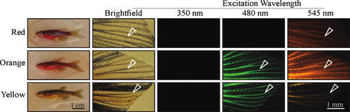
Patterns of fluorescence are consistent with orange GloFish carrying both the GloRFP and GloYFP transgenes. Progeny from Cross 2 in Figure 2 were assayed for emission of fluorescence after excitation by different wavelengths of light. Each row contains images of the same fish. The first column has images of the whole fish, and the remaining columns have images of the surgically removed caudal fins. The prominent structures are the fin rays (open white arrowheads). The red fish, consistent with the properties of RFP, fluoresces weakly under blue light and strongly under green light. In contrast to the yellow fish, the fluorescence is present in both the fin rays and the mesenchymal tissues in between. The yellow fish, consistent with the known properties of YFP, fluoresces strongly under blue light (480 nm) and only weakly under green light (545 nm). Fluorescence under both wavelengths is largely localized to the fin rays. The orange fish fluoresces strongly when excited by blue and green light, and fluorescence is present in the fin rays and in between. None of the GloFish fluoresce under UV light (350 nm). Analysis of all of the progeny demonstrated that fish with the same color under white, room light also have matching fluorescence patterns (Supplementary Material 7). All images are lateral views with anterior to the left. |