
Fig. 1
- ID
- ZDB-FIG-121217-28
- Publication
- Nguyen-Chi et al., 2012 - Morphogenesis and Cell Fate Determination within the Adaxial Cell Equivalence Group of the Zebrafish Myotome
- Other Figures
- All Figure Page
- Back to All Figure Page
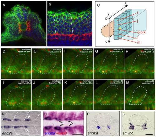
Distinct muscle precursor populations within the adaxial compartment. (A-C) The adaxial cell compartment. (A) Cross-section and (B) single confocal scan, dorsal view of a newly formed somite where the adaxial cells express the sMyHC (red). Nuclei and membranes are also marked with DAPI (blue) and membrane-bound GFP (green). (C–M) Restrospective fate map of the adaxial cell compartment. Adaxial cell behaviors occurring during the first phase of differentiation were analyzed in time lapse using a membrane-bound GFP (green) and a nuclear localized mCherry (red). The anterior-most adaxial cells in the dorso-ventral midline (yellow dot) are the first to differentiate and elongate. Adaxial cells above and below remain undifferentiated. The positions of individual confocal planes on the dorso-ventral axis are represented in (C). (N-Q) eng2a expression initiates in anterior adaxial cells at the dorso ventral midline of the myotome. 10-somite stage embryos on which in situ hybridization (ISH) for eng2a was performed alone (Blue, N, P) or in combination with myoD (Red, O,) that marks the posterior somitic region and the adaxial cells. Arrows indicate eng2a expression in the anterior adaxial cells. (Q) In situ hybridisation for smyhc demonstrates the location of the adaxial cells. (N, O) dorsal view, anterior toward the left, (P, Q) cross sections. Scale bar 50 μM. |
| Genes: | |
|---|---|
| Fish: | |
| Anatomical Terms: | |
| Stage: | 10-13 somites |