|
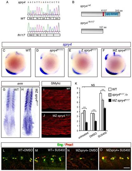
sprouty4 loss-of-function leads to the reduction of MP formation. (A) Sequencing of spry4WT and spry4fh117 confirms the single A to T mutation in position 469 of the spry4 ORF, resulting in the premature stop codon. (B) Predicted peptide arising from spry4WT and spry4fh117. The mutation results in a truncated protein of 157 amino acids (K157 in red is replaced by a stop codon) lacking the conserved cysteine rich domain [31] also called the spry domain (blue). This domain has been involved in RTK inhibition in other systems. (C–F) spry4 expression at 10-somite in WT, spry4fh117/+, Z spry4fh117 and MZ spry4fh117 embryos as determined by in situ hybridization. Whole mounted embryos, lateral view. (G–H) Erm expression in 9-somite WT and MZ spry4fh117 embryos. Flat mounted embryos, anterior toward the top. (I–J) sMyhc expression (red) in the somites of (I) WT and (J) MZ spry4fh117 embryos at 1 dpf. Maximal projection of multiple confocal scans, lateral view. (K) Graphic representation of the number of MPs per somite of 1 dpf embryos in different conditions: WT, spry4fh117/+, and MZ spry4fh117 untreated embryos or after DMSO or SU5402 treatments, values = means, error bars = s.e.m, ***p<0.001. (L–O) Engrailed (green) and Prox1 (red) expression in the somites of 1 dpf WT or MZ spry4fh117 embryos, after DMSO or SU5402 treatments, scale bar = 50 μm.
|