
Fig. 1
- ID
- ZDB-FIG-120914-13
- Publication
- Watkins et al., 2012 - High resolution imaging of vascular function in zebrafish
- Other Figures
- All Figure Page
- Back to All Figure Page
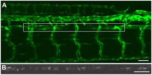
Intravital imaging of vascular dynamics in a 5 dpf zebrafish larva. An extended depth of focus projection (dorsal upwards, anterior left) of the vasculature of a Tg(kdrl:GFP)la116;Tg(gata1:dsRed)sd2 embryo with the fluorescence defining the endothelium (A). The outlined box shows the area of the aorta used for the time-based imaging of blood flow velocity and vessel diameter. The lower panel (B) shows a representative image from the same fish of the DsRed labeled erythrocytes within the dorsal aorta. This image was collected with a 2 ms exposure. With these settings it was readily possible to visualize the red blood cells flowing through the dorsal aorta over time. (Scale Bar = 100 microns). |