
Fig. 2
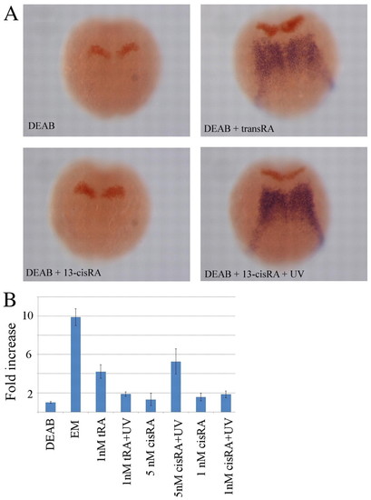
Response of vhnf1 to transient exposure to all-trans RA and 13-cis RA. Zebrafish embryos were incubated from sphere stage in 10 μM DEAB (except for the control incubated in embryo medium, EM), exposed at 90% epiboly for 5 minutes to various concentrations of all-trans RA (tRA) and 13-cis RA (cisRA) and UV illuminated (for 1 minute) or not. (A) In situ hybridization at the 1- to 2-somite stage for vhnf1 (blue) and krox20 (red) in embryos incubated in DEAB and 5 nM 13-cis RA (no vhnf1 is visible) or DEAB plus 1 nM all-trans RA and DEAB plus 5 nM 13-cis RA plus 1-minute UV illumination (rescue of vhnf1 expression is visible). (B) Quantification of the expression of vhnf1 by RT-qPCR in various conditions. Note that the expression of vhnf1 in 1 nM all-trans RA is similar to that in 5 nM 13-cis RA plus UV. Also, the expression of vhnf1 is similar in 1 nM all-trans RA plus UV and 1 nM 13-cis RA plus UV, as expected because in both cases the concentration of all-trans RA after UV illumination is the same. Error bars indicate s.e.m. from five experiments (for each experiment, RT-qPCR for the various genes was performed in triplicate). |