
FIGURE
Fig. S17
- ID
- ZDB-FIG-120824-6
- Publication
- Chen et al., 2012 - Haemodynamics-driven developmental pruning of brain vasculature in zebrafish
- Other Figures
- All Figure Page
- Back to All Figure Page
Fig. S17
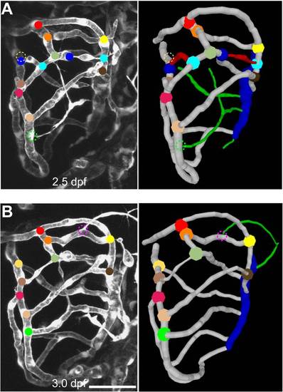
Method for tracing fate of each vessel segment in the midbrain. (A and B) Projected confocal images (left) and 3-D reconstructions (right) of half midbrain vasculature in a Tg(kdrl:eGFP) zebrafish larvae at 2.5 dpf (A) or 3 dpf (B). Colored balls mark different branch points. The dashed circle marks the site at which a branch point will appear at the next imaging time point. The corresponding movies of the 3-D rotation centerlines are shown in Video S7. Green, newly formed segments; red, pruning segments; blue, CVP. Scale, 50 μm. |
Expression Data
Expression Detail
Antibody Labeling
Phenotype Data
Phenotype Detail
Acknowledgments
This image is the copyrighted work of the attributed author or publisher, and
ZFIN has permission only to display this image to its users.
Additional permissions should be obtained from the applicable author or publisher of the image.
Full text @ PLoS Biol.