|
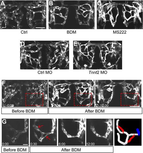
Effects of heartbeat suppression on vessel pruning. (A–C) Projected images of zebrafish larval midbrain vasculature at 50 hpf. Larvae were treated with normal solution (Ctrl, A), MS222 (tricaine, 0.66 mg/ml; B), or 2,3-butanedione-2-monoxime (20 mM, BDM; C) from 48 hpf and imaged at 50 hpf. (D and E) Projected images of zebrafish larval midbrain vasculature at 2 dpf. Larvae were microinjected with 4 ng control morpholino (Ctrl MO; D) or 4 ng Tnnt2 MO (E). Scale, 50 μm. (F–G) Time-lapse serial imaging showing BDM-induced vessel pruning. The morphology of vessels in the whole midbrain (F) and highlighted area (G) were shown before BDM treatment and after the onset of BDM treatment. BDM was bath-applied during 2–2.5 dpf and imaging was performed during this period. The regressed segments are pointed by the red arrows in the real images or marked in red in the 3-D reconstruction (G). Time, hour:minute. Scales, 50 μm in (F) and 20 μm in (G).
|