
Fig. 4
- ID
- ZDB-FIG-120814-26
- Publication
- Ellertsdottir et al., 2012 - Developmental Role of Zebrafish Protease-Activated Receptor 1 (PAR1) in the Cardio-Vascular System
- Other Figures
- All Figure Page
- Back to All Figure Page
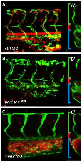
par1 knockdown impairs vascular remodeling in the cardinal vein. (A–B) Lateral views of double transgenic embryos (Tg(kdrl:EGFP)s843;Tg(gata1:dsRed)sd2) at 35 hpf. (A) The intersegmental vessels are connected to the CV and the one intersegmental artery (most posterior) seen in the figure has blood flowing from the dorsal aorta. (A2) In the mid-cross section, the intersegmental artery is visible (green bracket), the dorsal aorta can be seen as one tube (red bracket), and the CV is seen as two tubes (blue bracket). (B) par1 morphant lateral view. ISVs are lumenized (asterisk) (B2) In cross section, the CV tube does not show a defined number. Due to mislaid vein remodeling the tube is disorganized, cell clusters are apparent instead of a round tube. (C) The tnnt2 knockdown shows no mature inflation of the dorsal aorta and only a minor primary lumen in the ISV. The CV is bulged and full of blood cells in this region. (C′) The small dorsal aorta is a clear difference to the par1 knockdown, additionally to the lack of lumen in the ISVs and blood cells always clogging in the region of origin, PBI. |
| Fish: | |
|---|---|
| Knockdown Reagents: | |
| Observed In: | |
| Stage: | Prim-15 |