FIGURE

Fig. 2

ID
ZDB-FIG-120810-56
Publication
Lee et al., 2012 - Disruption of the folate pathway in zebrafish causes developmental defects
Other Figures
All Figure Page
Back to All Figure Page
Fig. 2

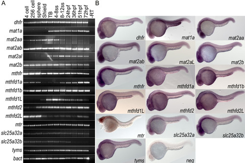
Wild type expression data for folate pathway transcripts. A) RT-PCR analysis of 11 embryonic stages within the first 72 hours of development indicates that genes involved in the folate pathway are generally maternally loaded transcripts that are expressed throughout early development. B) in situ hybridization for these 16 genes at 24 hpf shows that many are globally expressed with enrichment in anterior neural tissues including tectonic and retinal staining. Specific somatic staining is seen in mat1a, mat2aa, mat2ab, and mthfr. YSL staining is also observed in mat1a and mthfd1b. Embryos in lateral view.

Expression Data
Genes:
Fish:
Anatomical Terms:
Stage Range: 4-cell to Protruding-mouth

Expression Detail
Antibody Labeling
Phenotype Data

Phenotype Detail
Acknowledgments
This image is the copyrighted work of the attributed author or publisher, and ZFIN has permission only to display this image to its users. Additional permissions should be obtained from the applicable author or publisher of the image. Full text @ BMC Dev. Biol.