|
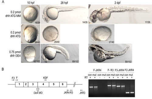
dhfrmorpholino injection phenocopies methotrexate treatment defects. A) Epiboly in dhfr ATG morphants is significantly delayed at 10 hpf as compared to controls. By 28 hpf severe dhfr ATG morphants are developmentally arrested with a failure to complete epiboly. Intron-exon splice morphants complete epiboly but exhibit shortened anterior-posterior axis and heart defects. Embryos in lateral view. B) Diagram of dhfr mRNA, i3e4 splice blocking morpholino, and primers used for RT-PCR (sequences supplied in Additional file 2: Table S2). cDNA generated from embryos at 6 hpf demonstrate splicing defects in dhfr transcripts in embryos treated with morpholino (mut). Control injected embryos (con) have only expected band sizes.
|