
Fig. 9
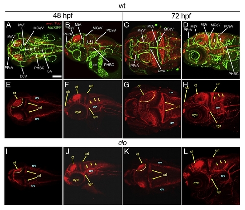
The axonal scaffolds develop largely independently of the vasculature. A-L, Maximum intensity confocal projections of immuno-fluorescently stained embryos carrying the endothelial-specific reporter Tg(kdrl:GFP)1a116. Endothelium, green (GFP). Axonal tracks, red (acetylated tubulin). Ages (hpf) indicated above. A-H, wild type (wt). I-L, clo (only the red channel is shown for simplicity). Abbreviations (see Table 1): vasculature, white (apostrophe, right side); axonal scafolds, yellow. Small white arrows, CtAs. Yellow dotted lines (E, I, G, K), dorsal limit of the optic tectum. Small yellow arrows, neurons with axonal commissures. A, E, I, C, G, K, Dorsal views. Anterior, left. Left side, bottom. B, F, J, D, H, L, Left lateral views. Anterior, left. Dorsal, top. Scale bar (A), 200 μm. |
| Gene: | |
|---|---|
| Antibody: | |
| Fish: | |
| Anatomical Terms: | |
| Stage Range: | Long-pec to Protruding-mouth |
| Fish: | |
|---|---|
| Observed In: | |
| Stage Range: | Long-pec to Protruding-mouth |
Reprinted from Developmental Biology, 357(1), Ulrich, F., Ma, L.H., Baker, R.G., and Torres-Vazquez, J., Neurovascular development in the embryonic zebrafish hindbrain, 134-51, Copyright (2011) with permission from Elsevier. Full text @ Dev. Biol.