
Fig. 8
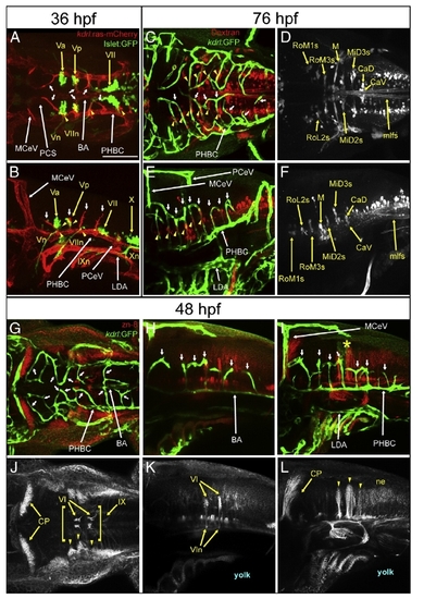
Anatomical relationship between hindbrain CtAs with branchiomotor neurons (BMNs), reticulospinal neurons (RSNs) and zn-8 positive neurons and axons. A-B, Maximum intensity confocal projections of immuno-fluorescently stained 36 hpf Tg(kdrl:ras-mCherry)s896; Tg(Islet1:GFP) embryos. Endothelium, red (mCherry). BMNs and their efferents, green (GFP). A, Dorsal view (ventral level). Anterior, left. Left side, bottom. B, Left lateral view. Anterior, left. Dorsal, up. C-F, Maximum intensity confocal projections of immunofluorescently stained 76 hpf embryos carrying the endothelial-specific reporter Tg(kdrl:GFP)1a116. RSNs retrogradely labeled with fluorescent Dextran. Endothelium, green (GFP). RSNs, red (C,E) or white (D,F). Abbreviations (see Table 1): vasculature, white. Neurons, yellow. Yellow arrowheads indicate regions where CtAs and RSNs are in close proximity to each other (C,E). C,D, Dorsal views. Anterior, left. Left side, bottom. E,F, Left lateral views. Anterior, left. Dorsal, up. G-L, Maximum intensity confocal projections of immunofluorescently stained 48 hpf embryos carrying the endothelial reporter Tg(kdrl:GFP)1a116. Endothelium, green (GFP). zn-8 positive (DM-GRASP/Neurolin) neurons and axons, red (G-I) or white (J-L). G,J, dorsal views. Anterior, left. Left side, bottom. H-I,K-L, left lateral views. Anterior, left. Dorsal, top. Abbreviations (see Table 1): vasculature, white; nuclei, nerves and commissures, yellow. Small white arrows, CtAs. Yellow arrowheads, regions where CtAs are in close proximity to nuclei or corresponding axons/neurites or prominent clusters of neurons in the neuroectoderm′s dorso-lateral region. Yellow brackets in (J), ventral axonal commissures. Yellow asterisk, r5 GFP-positive neuroepithelial signal from the Tg(kdrl:GFP)1a116 reporter. In (J-L), the abducens (VI) and glosso-pharyngeal (IX) motor neurons, the abducens nerves (VIn, arrows point to their hindbrain exit points) and the cerebellar plate (CP) are labeled. Scale bar (A), 100 μm. |
| Genes: | |
|---|---|
| Antibody: | |
| Fish: | |
| Anatomical Terms: | |
| Stage Range: | Prim-25 to Protruding-mouth |
Reprinted from Developmental Biology, 357(1), Ulrich, F., Ma, L.H., Baker, R.G., and Torres-Vazquez, J., Neurovascular development in the embryonic zebrafish hindbrain, 134-51, Copyright (2011) with permission from Elsevier. Full text @ Dev. Biol.