
Fig. 4
- ID
- ZDB-FIG-120611-5
- Publication
- Chen et al., 2012 - Heterogeneity across the dorso-ventral axis in zebrafish EVL is regulated by a novel module consisting of sox, snail1a and max genes
- Other Figures
- All Figure Page
- Back to All Figure Page
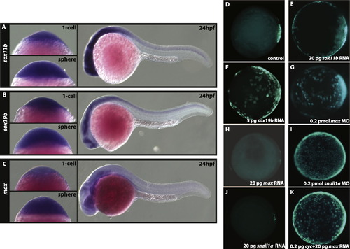
Expression analyses of sox11b, sox19b, and max. In situ hybridization of sox11b (A), sox19b (B), and max (C) at 1-cell, sphere, and 24hpf stages. All three genes are maternally contributed, ubiquitously expressed at sphere stage, and expressed in the CNS at 24 hpf (A–C). (D–K) Animal pole view of GFP expression in Tg (miniCrestin:Gal4, UAS:GFP) embryos at shield stage. In control embryos at the shield stage (D) GFP expression is restricted to the dorsal domain. This domain is expanded in sox11b (E), sox19b (F) over-expressing and max deficient (G) embryos. The over-expression of max (H) represses transgene expression in the dEVL. Similar to max, snail1a deficient embryos exhibit expansion (I) whereas over-expressing snail1a embryos (J) show reduction in the dEVL domain. The dEVL remains expanded in cyc and max mRNA co-injected embryos (K) indicating that Nodal signalling is sufficient to regulate dEVL formation. |
| Genes: | |
|---|---|
| Fish: | |
| Knockdown Reagents: | |
| Anatomical Terms: | |
| Stage Range: | 1-cell to Prim-5 |
Reprinted from Mechanisms of Development, 129(1-4), Chen, Y.Y., Harris, M.P., Levesque, M.P., Nüsslein-Volhard, C., and Sonawane, M., Heterogeneity across the dorso-ventral axis in zebrafish EVL is regulated by a novel module consisting of sox, snail1a and max genes, 13-23, Copyright (2012) with permission from Elsevier. Full text @ Mech. Dev.