
Fig. 6
- ID
- ZDB-FIG-120525-35
- Publication
- Jia et al., 2012 - Protein Phosphatase 4 Cooperates with Smads to Promote BMP Signaling in Dorsoventral Patterning of Zebrafish Embryos
- Other Figures
- All Figure Page
- Back to All Figure Page
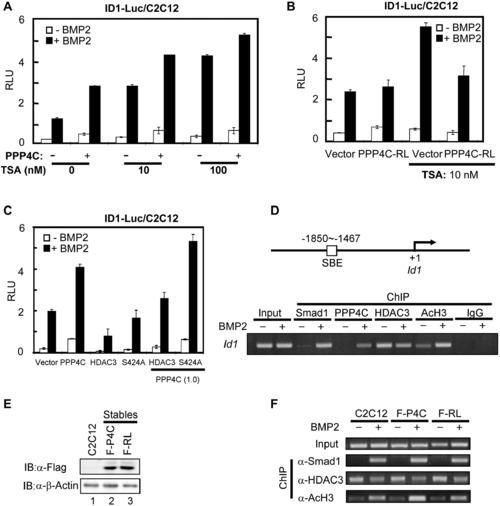
PPP4C Potentiating BMP Signaling Requires Its Phosphatase Activity to Antagonize HDAC3 Activity (A) PPP4C and HDAC inhibitor TSA similarly enhanced BMP-induced ID1-Luc reporter activity. Plasmids encoding PPP4C and ID1-Luc reporter were transfected into C2C12 cells. The luciferase activity was measured after cells were treated with or without BMP2 for 20 hr in the presence of vehicle or TSA at 10 or 100 nM. (B) Upregulation of BMP signaling by TSA treatment was blocked by PPP4C-RL (PPP4C-dead mutant). C2C12 cells transfected with plasmids encoding PPP4C-RL and ID1-Luc reporter were subjected to luciferase assays. (C) PPP4C enhanced BMP-induced ID1 promoter activity through antagonizing HDAC3 activity. C2C12 cells transfected with a combination of indicated plasmids were subjected to luciferase assays. In the parentheses is the amount of indicated plasmids (μg/4 well). S424A, an HDAC3 mutant that is refractory to PPP4C activity. (D) ChIP analysis of Id1 promoter occupancy by Smad1, Ppp4c, Hdac3, and AcH3 in response to BMP2 stimulation. C2C12 cells were treated with or without BMP2 for 2 hr; cell lysates were subjected to ChIP assays using indicated antibodies. Anti-Rabbit IgG ChIP served as negative control. Bottom view shows that Id1 promoters were detected by gel electrophoresis after regular PCR; top view is a diagram showing the PCR-amplified region on the Id1 promoter. (E) Comparable ectopic expression level of PPP4C and PPP4C-RL in C2C12 stables. Whole-cell lysates from C2C12 cells stably expressing Flag-PPP4C (F-P4C) or Flag-PPP4C-RL (F-RL) as well as the parental C2C12 cells were analyzed by western blot with indicated antibodies. β-Actin blot served as loading control. (F) PPP4C enhanced histone acetylation at the Id1 promoter in phosphatase activity-dependent manner. ChIP analysis in C2C12 stables and parental C2C12 cells was similarly conducted as in (D). Data are presented with mean ± SD. See also Figure S5. |
Reprinted from Developmental Cell, 22(5), Jia, S., Dai, F., Wu, D., Lin, X., Xing, C., Xue, Y., Wang, Y., Xiao, M., Wu, W., Feng, X.H., and Meng, A., Protein Phosphatase 4 Cooperates with Smads to Promote BMP Signaling in Dorsoventral Patterning of Zebrafish Embryos, 1065-1078, Copyright (2012) with permission from Elsevier. Full text @ Dev. Cell