
Fig. 5
- ID
- ZDB-FIG-120525-34
- Publication
- Jia et al., 2012 - Protein Phosphatase 4 Cooperates with Smads to Promote BMP Signaling in Dorsoventral Patterning of Zebrafish Embryos
- Other Figures
- All Figure Page
- Back to All Figure Page
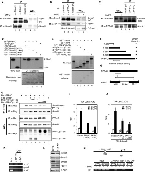
Ppp4c Is a Direct Interacting Partner of Smad1 and Smad5 (A–C) Ppp4c interacted with Smad1 and Smad5 at endogenous level in C2C12 cells. The indicated proteins were immunoprecipitated or detected using corresponding antibodies. WCL, whole-cell lysate; IP, immunoprecipitation; IB, immunoblot. (D) PPP4C directly interacted with MH1 and MH2 domain of Smad1. In-vitro-synthesized full-length PPP4C and control GFPs were S35 labeled and incubated with GST protein fused with WT Smad1 (GST-Smad1) or mutant forms. Retrieved proteins were detected by autoradiography (top); Coomassie blue staining demonstrated the amount of used GST proteins (bottom). (E and F) Smad1 directly interacted with PPP4C. In vitro GST-pull-down assays were similarly conducted as in (D). The results are summarized in a schematic view in (F). (G) A schematic view of PPP4C-Smad1 interactions. (H) PPP4C deletion mutants competed with PPP4C for binding to Smad1. CoIP analysis of Smad1-PPP4C interaction in the presence of coexpressed PPP4C truncation mutants was conducted in HEK293T cells. (I and J) PPP4C truncation mutants inhibited BMP2-induced ID1 reporter activity (I) and FR-Luc reporter activity (J). (K) Endogenous Ppp4c in zebrafish embryos bound to the promoter of ventral genes eve1, gata2, and foxi1. Zebrafish embryos at the shield stage were subjected to ChIP assays using anti-PPP4C antibody, followed by detection of endogenous promoter DNA by PCR. Anti-Rabbit-IgG ChIP served as negative control. (L) Effective depletion of Smad1/Smad8 in C2C12 stables. Whole-cell lysates from C2C12 cells stably expressing pSRG vector (CTRL) or shRNA-Smad1/Smad8 (S1/8-KD) were analyzed by western blot with indicated antibodies with β-actin blot as loading control. (M) BMP2 induced Ppp4c binding to the endogenous Id1 promoter. On the top is the diagram showing the PCR-amplified region on endogenous Id1 promoter. On the bottom is the ChIP result. Depletion of Smad1/Smad8 inhibited BMP2-induced Ppp4c binding to the Id1 promoter in C2C12 cells. Anti-Rabbit IgG ChIP served as negative control. Data are presented with mean ± SD. |
Reprinted from Developmental Cell, 22(5), Jia, S., Dai, F., Wu, D., Lin, X., Xing, C., Xue, Y., Wang, Y., Xiao, M., Wu, W., Feng, X.H., and Meng, A., Protein Phosphatase 4 Cooperates with Smads to Promote BMP Signaling in Dorsoventral Patterning of Zebrafish Embryos, 1065-1078, Copyright (2012) with permission from Elsevier. Full text @ Dev. Cell