
Fig. 4
- ID
- ZDB-FIG-120525-33
- Publication
- Jia et al., 2012 - Protein Phosphatase 4 Cooperates with Smads to Promote BMP Signaling in Dorsoventral Patterning of Zebrafish Embryos
- Other Figures
- All Figure Page
- Back to All Figure Page
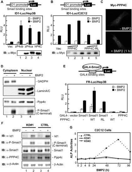
PPP4C Enhances BMP Signaling in Mammalian Cells (A) Zebrafish and human PPP4C enhanced the BMP2-induced ID1-Luc reporter expression. Indicated plasmids were cotransfected with ID1-Luc into Hep3B cells. The luciferase activity was measured 20 hr after BMP2 stimulation. The expression levels of transfected plasmids are shown in order in the lower panel. IB, immunoblot; RLU, relative luciferase activity; Vec, control vector pRK5; zP4ca and zP4cb, zebrafish Ppp4ca and Ppp4cb; hP4C, human PPP4C. (B) Human PPP4C required its phosphatase activity to enhance ID1-Luc reporter expression. PPP4C-RL is a phosphatase-dead mutant of human PPP4C. The expression levels of transfected plasmids are shown in order in the lower panel. (C) Overexpressed Myc-PPP4C in C2C12 cells was visualized by indirect immunofluorescence with or without BMP2 stimulation for 1 hr. (D) Endogenous Ppp4c was detected by western blot in both cytoplasmic and nuclear fractions of C2C12 cells. GADPH and LaminA/C served as the cytoplasmic and nuclear protein loading controls, respectively. C-terminal phosphorylation of Smad1 was detected with an anti-Phospho-Smad1 antibody (Cell Signaling Technology). (E) PPP4C enhanced the transcriptional activity from a Smad1-occupied promoter. FR-Luc reporter was transfected into Hep3B cells together with the combinations of GAL4-Smad1 (GAL4-S1) and Myc-tagged PPP4C (WT) or mutant PPP4C-RL (RL). The luciferase activity was measured after cells were treated with or without BMP2 for 20 hr. (F) Ppp4c depletion in C2C12 cells inhibited BMP-induced expression of endogenous Id1. Cell lysates were prepared from Ppp4c-KD#1 and control (CTRL) C2C12 cells stimulated with BMP2 for 2 hr. The expression levels of various proteins were examined by western blot. β-Actin served as the loading control. (G) Ppp4c depletion in C2C12 cells inhibited BMP2-induced osteoblastic differentiation. C2C12 cells were treated with BMP2 for up to 4 days to induce osteoblastic marker expression. Cell lysates were harvested at indicated time points and subjected to analysis of ALP activity. The time course of ALP activity was plotted. Data are presented with mean ± SD. See also Figure S4. |
Reprinted from Developmental Cell, 22(5), Jia, S., Dai, F., Wu, D., Lin, X., Xing, C., Xue, Y., Wang, Y., Xiao, M., Wu, W., Feng, X.H., and Meng, A., Protein Phosphatase 4 Cooperates with Smads to Promote BMP Signaling in Dorsoventral Patterning of Zebrafish Embryos, 1065-1078, Copyright (2012) with permission from Elsevier. Full text @ Dev. Cell