
Fig. 2
- ID
- ZDB-FIG-120508-45
- Publication
- Zhang et al., 2012 - The Expression of irx7 in the Inner Nuclear Layer of Zebrafish Retina Is Essential for a Proper Retinal Development and Lamination
- Other Figures
- All Figure Page
- Back to All Figure Page
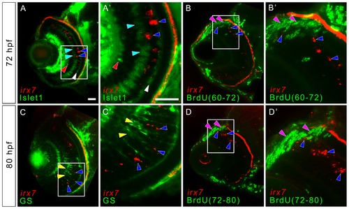
Irx7 expresses in non-proliferative cells that are likely to be undifferentiated precursors in mature retina. To detect for the co-localization of irx7 and ACs, BCs, HCs, MCs and proliferative cells in the more differentiated part of the WT retina, in situ hybridization of irx7 was conducted in conjunction with immunostaining of anti-Islet1 for ACs, BCs and HCs, anti-GS for MCs and anti-BrdU for proliferative cells. (A) irx7 in situ hybridization with anti-Islet1 immunostaining at 72 hpf. The blue arrowheads indicate the irx7+ cells (red colour) in the retina, while the red, cyan and white arrowheads indicate ACs, BCs and HCs respectively (all in green colour). (A2) The magnified view of the white box in (A). (B and D) The retina of embryos treated with BrdU from 60 to 72 hpf and 72 to 80 hpf respectively. The blue arrowheads indicate irx7+ cells (red colour), while the pink arrowheads indicate BrdU+ cells (green colour). (B2 and D2) The magnified view of the white box in (B and D) respectively. (C) irx7 in situ hybridization with anti-GS immunostaining at 80 hpf. The blue arrowheads indicate the irx7+ cells (red colour), while the yellow arrowheads indicate MCs (green colour). (C2) The magnified view of the white box in (C). Lateral is to the left and dorsal is up for all sections. Note that the RPE layer also showed red fluorescence but that was not a real signal. It was an artifact of the pigmentation in RPE. Since the images of the in situ hybridization were inverted before combining with the fluorescent images obtained from the immunostaining, the darker pigment in RPE, as well as the intense in situ colour, would appear as signal in this transformation. Scale bars = 20 μm. |
| Gene: | |
|---|---|
| Antibodies: | |
| Fish: | |
| Anatomical Terms: | |
| Stage: | Protruding-mouth |