
Fig. 7
- ID
- ZDB-FIG-120508-50
- Publication
- Zhang et al., 2012 - The Expression of irx7 in the Inner Nuclear Layer of Zebrafish Retina Is Essential for a Proper Retinal Development and Lamination
- Other Figures
- All Figure Page
- Back to All Figure Page
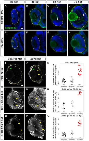
Irx7 knockdown does not induce apoptosis but can delay cell cycle withdrawal. Immunostaining of embryos injected with 10 ng of control MO or irx7SMO were conducted with anti-active caspase3 antibody (green colour) at 28, 36, 52 and 72 hpf. The nuclei were counterstained with DAPI (blue colour). In both controls (A–D) and irx7SMO morphants (E–H), only a few active caspase3+ cells (white arrowheads) were detected in some retinas at all stages. All cells that showed a positive anti-active caspase3 signal had the characteristic cell shrinkage and rounded morphology, as shown by the DAPI staining. While all active caspase3- cells looked healthy. Mitotic cells were detected by anti-PH3 in the retinas of controls (I) and morphants (J) at 72 hpf. PH3+ cells in the MZ and in the ectopic apical retina are indicated by green and yellow arrowheads respectively. (K) A stripchart of the number of PH3+ cell per retinal area in uninjected embryos, controls and morphants. Retinal cells that had gone through S-phase in controls and morphants were also detected by BrdU incorporation from 36 to 52 hpf (L and M) and from 52 to 72 hpf (O and P). (N and Q) The corresponding stripcharts of BrdU+ area per retinal area in the uninjected embryos, controls and morphants. The asterisks in all stripcharts represent the median of each group. Lateral is to the left and dorsal is up for all sections. The retinal region in these samples is highlighted by a dotted yellow line. Scale bar = 20 μm. |
| Fish: | |
|---|---|
| Knockdown Reagent: | |
| Observed In: | |
| Stage Range: | Prim-5 to Protruding-mouth |