
Fig. 5
- ID
- ZDB-FIG-120405-76
- Publication
- Behra et al., 2012 - Transcriptional signature of accessory cells in the lateral line, using the Tnk1bp1:EGFP transgenic zebrafish line
- Other Figures
- All Figure Page
- Back to All Figure Page
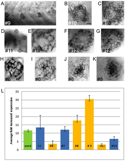
WISH and q-RT-PCR on novel genes enriched in accessory cells A to K. Whole mount in situ hybridizations (WISH) in 3 and 5 dpf larvae A ring like staining is visible, because hybridization is excluded from centrally located hair cells and only found in peripheral accessory cells for gene #9, 10, 11 and 12 (A to G). In #6 and 8 (H to K), the hybridization is present in the whole neuromast, but appears much stronger in centrally located hair cells. A. WISH against #9 in 3 three neuromasts in a 3 dpf larva (A). B and C. WISH against #10 in a neuromast in a 3 dpf (B) and a 5 dpf larva (C). D and E. WISH against #11 in a portion of the 5 dpf larval head showing 4 neuromasts (D) and of a neuromast in a 3 dpf larva (E). F and G. WISH against #12, in two neuromasts in 5 dpf larvae (F and G). H and I. WISH against #6 in a neuromast in a 3 dpf (H) and in a 5 dpf larvae (I). J and K. WISH against #8 in two neuromasts in 5 dpf larva (J and K). - is 50 microns in A and D, and 5 microns in B. L. Average fold increased expression of genes determined by qRT-PCR. The tnks1bp1 gene is in green. Genes showing an expression pattern by WISH (in A to Q) are in yellow the others are in blue. Error bars represent standard deviation. |
| Genes: | |
|---|---|
| Fish: | |
| Anatomical Terms: | |
| Stage Range: | Protruding-mouth to Day 5 |