
Fig. 1
- ID
- ZDB-FIG-120405-72
- Publication
- Behra et al., 2012 - Transcriptional signature of accessory cells in the lateral line, using the Tnk1bp1:EGFP transgenic zebrafish line
- Other Figures
- All Figure Page
- Back to All Figure Page
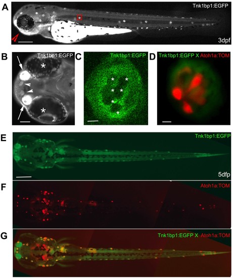
Live imaging of a 3 day old Tg (tnks1bp1:EGFP) embryo and a 5 day old Tg(tnks1bp1:EGFP) x Tg (atoh1a:dTOM) double transgenic larva. A. GFP is expressed in all of the neuromasts in the anterior (head) and posterior (trunk and tail) lateral line, as shown in a three day old embryo. B. Ventral view (red arrowhead in A) of the rostral head region of the embryo, showing a strong GFP expression in the olfactory epithelia (white arrows) between the eyes (white asterisks) and in the two more rostral neuromasts (white arrowheads). C. Magnification of a neuromast (red box in A) in a 3 dpf live embryo, showing GFP expression excluded from the six centrally located hair cells (white asterisks). D. Neuromast of a Tg(tnks1bp1:EGFP) × Tg(atoh1a:dTOM) 5 dpf double transgenic animal expressing GFP (green) in the accessory cells and TOMATO (red) in hair cells. E, F and G. Dorsal view of a 5 day dpf Tg(tnks1bp:GFP) × Tg(atoh1a:dTOM) double transgenic larva. - 200 microns in A and E, 40 microns in B, and 5 microns in C and D. |
| Genes: | |
|---|---|
| Fish: | |
| Anatomical Terms: | |
| Stage Range: | Protruding-mouth to Day 5 |