
Fig. 8
- ID
- ZDB-FIG-120315-59
- Publication
- Petrey et al., 2012 - Excessive activity of cathepsin K is associated with the cartilage defects in a zebrafish model for mucolipidosis II
- Other Figures
- All Figure Page
- Back to All Figure Page
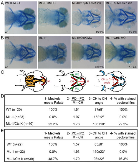
Inhibition of cathepsin K expression or activity results in correction of the ML-II cartilage morphogenesis defects. (A,B) Alcian blue stains of embryos at 4 dpf show that inhibition of either (A) cathepsin K activity using pharmacological agents or (B) expression by MO injection results in significant correction of multiple aspects of the craniofacial defects present in ML-II embryos. Percent values listed represent the number of embryos with these phenotypes. For the drug treatments (A), n=160 embryos in four experiments; for MO experiments (B), n=100 embryos in three experiments. (C) The degree of correction was quantified as follows: (1) whether Meckels (M) cartilage meets the palate (P), (2) the ‘shape’ of the jaw using the ratio of the distance between the palatoquadrate (PQ) bones over the distance from Meckels (M) cartilage to the ceratohyal (CH) bones, (3) the angle between the left and right ceratohyals (CH), and (4) whether the pectoral fins stained positively with Alcian blue. (D,E) The quantitation of the degree of cartilage correction following pharmacological inhibition of cathepsin K activity (D) and SB MO-inhibition of cathepsin K expression (E) are presented. |
| Fish: | |
|---|---|
| Knockdown Reagents: | |
| Observed In: | |
| Stage: | Day 4 |