
Fig. 5
- ID
- ZDB-FIG-120315-57
- Publication
- Petrey et al., 2012 - Excessive activity of cathepsin K is associated with the cartilage defects in a zebrafish model for mucolipidosis II
- Other Figures
- All Figure Page
- Back to All Figure Page
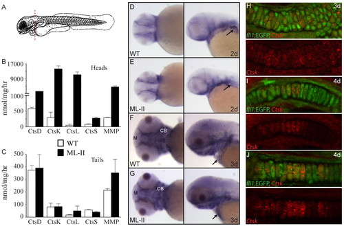
Cathepsin expression is enriched in the craniofacial skeleton of zebrafish embryos. (A–C) Analysis of enzyme activities in lysates of 3-dpf WT and ML-II embryos separated into heads and tails, as diagrammed in A (dashed line represents cut site) demonstrates that cathepsin activities are primarily increased in the head in ML-II embryos compared with WT (B) rather than in the tail (C); n=3. (D–G) In situ hybridization for cathepsin K expression shows mRNA enrichment in ventral tissues that generally correspond to the pharyngeal skeleton at 2 dpf (D,E) and 3 dpf (F,G) in WT and ML-II embryos. Arrows denote differences in the pattern of expression between WT and ML-II embryos. CB, ceratobranchials; M, Meckels cartilage. (H–J) Immunohistochemical stains for cathepsin K (red) on sections of 3 dpf (H) and 4 dpf (I,J) WT fli1a:EGFP embryos show that it is expressed in chondrocytes and peri-chondrial fibroblasts of the trabecular (H,J) and Meckels cartilages (I). |
| Gene: | |
|---|---|
| Antibody: | |
| Fish: | |
| Knockdown Reagent: | |
| Anatomical Terms: | |
| Stage Range: | Long-pec to Day 4 |