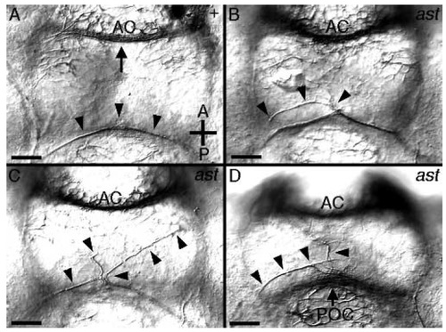
Anti-acetylated tubulin labeling of astray mutants. All panels show a ventral view of the ventral brain commissures at 36 hours of development. (A) In the wild type, the retinal ganglion cell axons grow near the POC (just dorsal to this focal plane). At 36 hours the first RGC axons (arrowheads) have reached the midline and fasciculated with their contralateral homologues. The anterior commissure is at the top of the photo (arrow) and the TPOC axons are lateral in a more dorsal focal plane. (D-D) Three examples of errors made at the midline by RGC growth cones in astray mutants. In all embryos, the POC has formed normally (most easily seen in D, arrow). RGC axons (arrowheads) grow abnormally on the ventral diencephalon. Instead of growing along their contralateral counterparts, RGC axons spread on the ventral diencephalon (B,D). In some cases axons from the two eyes clearly remain separate and appear to avoid each other (C). AC, anterior commissure; POC, postoptic commissure. Scale bars, 25 µm.
|
