
- Title
-
Zebrafish mutations affecting retinotectal axon pathfinding
- Authors
- Karlstrom, R.O., Trowe, T., Klostermann, S., Baier, H., Brand, M., Crawford, A.D., Grunewald, B., Haffter, P., Hoffman, H., Meyer, S.U., Muller, B.K., Richter, S., van Eeden, F.J., Nüsslein-Volhard, C., and Bonhoeffer, F.
- Source
- Full text @ Development

Retinotectal projections in the wild type and in mutants with ipsilateral retinotectal projections. Dorsal view, anterior to the left. Fish were stained with DAPI (blue fluorescence) to outline the tectal neuropil, which is surrounded by DNAcontaining cell bodies (see methods). Neuropilar regions are often difficult to see in these photographs and one neuropil is outlined in B. Black spots are melanophores in the skin (yellow arrowhead in B) (A) Diagram of an injected 5-day-old zebrafish. The left eye of each fish was injected with DiI (red) in the temporal/ventral quadrant and with DiO (green) in the nasal/dorsal quadrant except in D, E and F, where the dyes were reversed. Labeled retinal ganglion cell axons cross the brain and grow to the contralateral tectal lobe where they project topographically. (B) Wild-type projections, dorsal focal plane. On the contralateral tectal lobe, retinal ganglion cell axons project topographically with temporal/ventral RGC neurons sending axons to the anterior/dorsal tectum (arrowhead) and nasal/dorsal RGC neurons sending axons to the posterior/ventral tectum (arrow). (C) Wild-type projections, ventral focal plane; termination zones are now out of focus. Retinal ganglion cell axons leave the eye at the papilla and grow along the base of the diencephalon to the midline, where they cross completely at the optic chiasm. After the chiasm, axons sort into a dorsal (arrowhead) and a ventral (arrow) brachium and grow dorsoposteriorly to the contralateral tectal lobe. (D,E) Retinal ganglion cells project to the ipsilateral tectal lobe in belladonna mutants. Reverse dye injections. (D)Ventral focal plane, showing that the axons turn dorsal immediately after leaving the eye (arrow). (E) Dorsal focal plane showing normal topographic projections on the ipsilateral tectal lobe. (F) RGC axons also project to the ipsilateral tectal lobe in you-too mutants. (G,H) In detour mutants, RGC axons either project to the ipsilateral and contralateral tectal lobes in the same fish (G), or to the ipsilateral tectal lobe only (H). (G) Dorsal focal plane showing normal mapping of nasodorsal RGC axons (arrows) and ventral/temporal axons (arrowheads) from one eye on both tectal lobes. (H) In some dtr mutant fish, RGC axons grow to the midline (arrow) before turning back to the ipsilateral tectal lobe. (I) In blw mutants, RGC axons grow across the midline (arrow), then return to the ipsilateral tectal lobe. OP, olfactory placode; N, nasal; T, temporal. A, anterior; P, posterior; D, dorsal; V, ventral. Scale bars, 100 mm. PHENOTYPE:
|
|
ZFIN is incorporating published figure images and captions as part of an ongoing project. Figures from some publications have not yet been curated, or are not available for display because of copyright restrictions. PHENOTYPE:
|
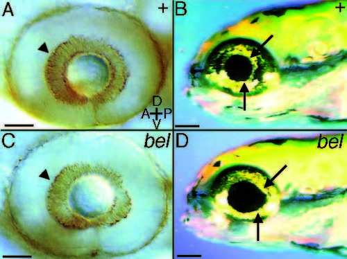
belladonna mutant eye phenotype. Wild-type (A) and belladonna (C) eyes at 48 hours labeled with the ZN-5 antibody to visualize retinal ganglion cell bodies. The retinal ganglion cells (arrowheads) and the entire eye appear normal at this age, thus the mutation does not appear to affect RGC differentiation. At 5 days iridophores and the pigmented epithelium are adjacent to the lens in the wild type (B). In belladonna mutants (D), gaps appear between the lens and some parts of the pigmented epithelium (arrow). The lens appears normal however. A, anterior; P, posterior; D, dorsal; V, ventral. Scale bars, 50 µm (A,C), 100 µm (B,D). |
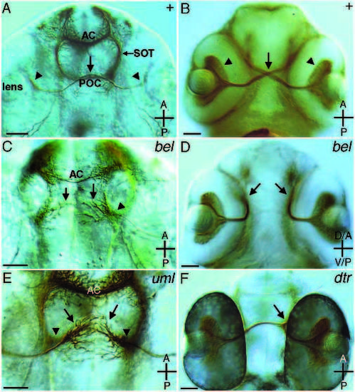
ZN-5 and anti-AT antibody labeling of wild type and mutants with ipsilateral retinotectal projections. (A, C and E) 36-hour embryos labeled with the anti-acetylated tubulin antibody that recognizes most axons. (B, D and F) 48-hour embryos labeled with the ZN-5 antibody that recognizes RGCs and their axons. (A) At 36 hours in the wild type, RGC axons have crossed the midline and grow along their contralateral counterparts (arrow). Arrowheads show retinal ganglion cell bodies. (B) At 48 hours in the wild type, the optic chiasm is well formed (arrow) and RGC axons extend to the contralateral tectal lobes. RGCs are positioned near the lens (arrowheads). (C) In belladonna mutants at 36 hours, the POC does not form (arrows), while the AC forms relatively normally. RGC axons remain near the ipsilateral eye (arrowhead). (D) In belladonna mutants at 48 hours, RGC axons project dorsally immediately after leaving the eye (arrows). (E) In some 36-hour umleitung mutant embryos, RGC axons grow toward the midline (arrows). The axons of the tract of the post-optic commissure remain lateral (arrowheads) and no POC forms. In other fish, RGC axons seem to turn dorsally immediately after leaving the eye (not shown). (F) In some detour mutant embryos, RGC axons from one eye project ipsilaterally (arrow), while those from the other eye project contralaterally. Axons can be followed to the tectal lobe on the right in this figure, while no axons project to the other tectal lobe. The result is that both eyes project to the same tectal lobe. AC, anterior commissure; POC, post-optic commissure; SOT, supraoptic tract. A, anterior; P, posterior; D, dorsal; V, ventral. Scale bars, 50 µm (A,B,D,F), 25 µm (C,E). |
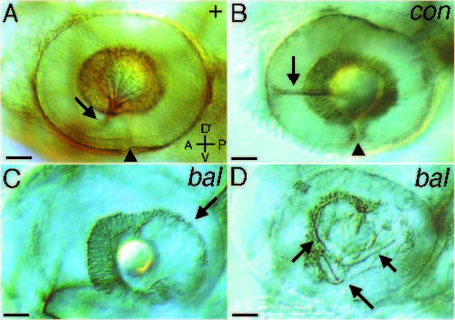
Pathfinding errors within the eye. Lateral views of eyes labeled with ZN-5 at 48 hours of development. Arrowheads mark the ventral fissure. (A) In the wild type, RGC axons aggregate in the ventral medial portion of the eye and exit at the optic nerve head (arrow). (B) In con mutants, RGC axons often do not exit the eye and instead grow along the equator of the eye in both anterior (arrow) and posterior (out of focus) directions. (C) RGC cell bodies do not surround the lens normally in bal mutants. In these fish temporal RGCs are not normally formed (arrow). Lateral focal plane. (D) Some bal mutants also have misrouted axons within the eye (arrows). These axons are disorganized within the eye, but do exit the eye and eventually project into the brain. Medial focal plane. A, anterior; P, posterior; D, dorsal; V, ventral. Scale bars, 50 µm. |
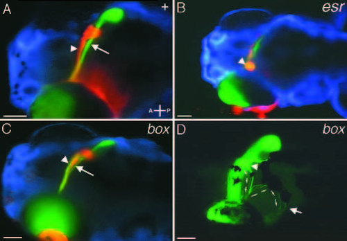
Retinotectal projections in mutants with reduced midline crossing and midline sorting errors. Ventral focal planes. (A) Wildtype projections showing ventral (arrow) and dorsal (arrowhead) brachia of the optic tract. (B) In esrom (and tilsit and tofu, not shown) mutants a large percentage of retinal ganglion cell axons fail to cross the midline. Instead they form an aggregate of fibers (arrowhead) just lateral to the papilla. (C) In boxer mutants, axons from dorsally located retinal ganglion cells do not sort correctly at the optic chiasm. Normally, all axons from dorsal RGCs follow the ventral brachium to the ventral side of the tectum. In boxer (and dackel, not shown), these axons split and follow both the ventral (arrow) brachium and the dorsal (arrowhead) brachium. Axons arriving at the wrong (dorsal) side of the tectum nonetheless find their appropriate ventral target region (see Trowe et al., 1996). (D) In some cases nasal/dorsal RGC axons that grow in the inappropriate (dorsal) brachium fail to enter the contralateral tectal lobe in boxer mutants (arrowhead). These axons (small arrows) project to the ipsilateral tectal lobe by crossing the dorsal midline and terminate in their appropriate retinotopic position (arrow). A, anterior; P, posterior. Scale bars, 100 µm. PHENOTYPE:
|
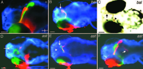
Retinotectal projections in mutants with anterior/posterior pathfinding errors. Ventral focal plane. (A) Wild-type projections. (B) In bashful mutants, axons project anteriorly into the forebrain, where they grow along the edge of the telencephalon (arrow) and occasionally make a complete circuit of the forebrain. (C) In some cases axons project to the ipsilateral tectal lobe in bashful. In these fish, RGC axons project both anteriorly (arrow) and turn (arrowhead) toward the ipsilateral tectal lobe (not seen in this focal plane). The DiI label was photoconverted to make the labeled RGC axons visible using DIC optics. (D-F) In astray mutants, axons grow aberrantly to one of three general destinations. After crossing the midline, axons either grow anteriorly into the anterior telencephalon (D, arrow), fan out toward the contralateral eye (E, arrow) and/or return to the ipsilateral side of the brain (E, arrowhead), or grow ventrally under the tectum to the hindbrain (F, arrow) (reverse dye fill). A majority of axons find their correct target region in the tectum in F. In these cases, axons fail to turn dorsally after crossing the midline and remain at the same dorsal/ventral level as the optic nerve. A, anterior; P, posterior. Scale bars, 100 µm. |
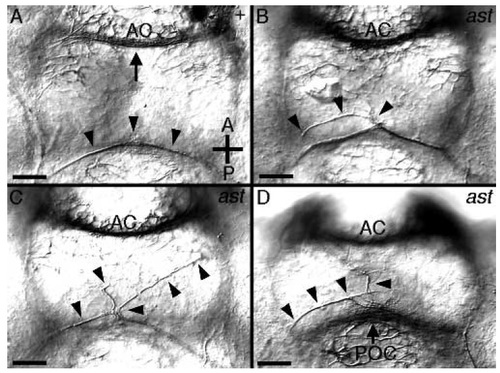
Anti-acetylated tubulin labeling of astray mutants. All panels show a ventral view of the ventral brain commissures at 36 hours of development. (A) In the wild type, the retinal ganglion cell axons grow near the POC (just dorsal to this focal plane). At 36 hours the first RGC axons (arrowheads) have reached the midline and fasciculated with their contralateral homologues. The anterior commissure is at the top of the photo (arrow) and the TPOC axons are lateral in a more dorsal focal plane. (D-D) Three examples of errors made at the midline by RGC growth cones in astray mutants. In all embryos, the POC has formed normally (most easily seen in D, arrow). RGC axons (arrowheads) grow abnormally on the ventral diencephalon. Instead of growing along their contralateral counterparts, RGC axons spread on the ventral diencephalon (B,D). In some cases axons from the two eyes clearly remain separate and appear to avoid each other (C). AC, anterior commissure; POC, postoptic commissure. Scale bars, 25 µm. |
|
Unillustrated author statements |