|
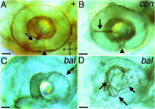
Pathfinding errors within the eye. Lateral views of eyes labeled with ZN-5 at 48 hours of development. Arrowheads mark the ventral fissure. (A) In the wild type, RGC axons aggregate in the ventral medial portion of the eye and exit at the optic nerve head (arrow). (B) In con mutants, RGC axons often do not exit the eye and instead grow along the equator of the eye in both anterior (arrow) and posterior (out of focus) directions. (C) RGC cell bodies do not surround the lens normally in bal mutants. In these fish temporal RGCs are not normally formed (arrow). Lateral focal plane. (D) Some bal mutants also have misrouted axons within the eye (arrows). These axons are disorganized within the eye, but do exit the eye and eventually project into the brain. Medial focal plane. A, anterior; P, posterior; D, dorsal; V, ventral. Scale bars, 50 µm.
|