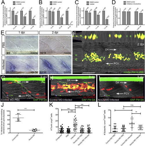
Demand-Driven Granulopoiesis and HSPC Expansion Are Dependent upon Nos2a Relative frequency of infected larvae demonstrating demand-driven granulopoiesis following treatment (from 6 hpi) with the NOS inhibiting drugs L-NAME (A), ODQ (B), 1400W (C), and S-Methyl-L-thiocitrulline (D), all relative to DMSO control treatment. Numbers represent numbers of larvae in each treatment group. (E) Expression of nos2a within the AGM of infected larvae, compared with PBS controls at 1 and 2 dpi. (F) Dual WMISH of nos2a and lyz within the AGM of infected larvae at 2 dpi. (G–I) DAF-FM DA detection of NO within control MO-injected PBS control, control MO-injected infected, and Nos2a MO-injected infected Tg(lyz:DsRED) larvae, respectively, at 2 dpi. Yellow and red asterisks mark lyz+/NO+ and lyz+/NO* cells, respectively. (J) Percentage of control MO- and Nos2a MO-injected Tg(lyz:DsRED) infected larvae demonstrating demand-driven granulopoiesis at 2 dpi. (K) Number of cmyb-expressing HSPCs, as detected by WMISH at 2 dpi, within the AGM compartment (as defined in Figure 2C) following indicated treatments. (L) Quantification of dual-labeled HSPCs within the subaortic space of 2 dpi Tg(runx1P2:EGFP)/Tg(kdrl:nls-mCherry) larvae following indicated treatments. Numbers represent frequency of embryos or larvae with displayed phenotype. All views, anterior to left. Scale bar, 50 μm in (F). Abbreviations: DA, dorsal aorta; PCV, posterior cardinal vein; n.s., not significant; **p < 0.01; ***p < 0.001. See also Figure S5.
|
