
FIGURE
Fig. S3
Fig. S3
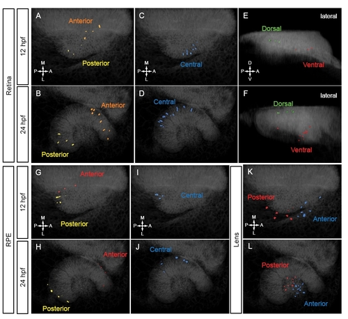
Clustering of tracked cells. Tracked cells were assigned to domains based on final position (24 hpf) and trajectory shape. Volume renderings of tracked nuclei at 12 (A,C,E,G,I,K) and 24 (B,D,F,H,J,L) hpf. (A-F) Retinal nuclear positions. (A,B) Anterior and posterior domains. Dorsal view. (C,D) Central domain. Dorsal view. (E,F) Dorsal and ventral domains. Lateral view. (G-J) RPE nuclear positions. (G,H) Anterior and posterior domains. Dorsal view. (I,J) Central domain. Dorsal view. (K,L) Lens nuclear positions. Anterior and posterior domains. |
Expression Data
Expression Detail
Antibody Labeling
Phenotype Data
Phenotype Detail
Acknowledgments
This image is the copyrighted work of the attributed author or publisher, and
ZFIN has permission only to display this image to its users.
Additional permissions should be obtained from the applicable author or publisher of the image.
Full text @ Development