
Fig. 6
- ID
- ZDB-FIG-111117-18
- Publication
- Xi et al., 2011 - Transgenic zebrafish expressing green fluorescent protein in dopaminergic neurons of the ventral diencephalon
- Other Figures
- All Figure Page
- Back to All Figure Page
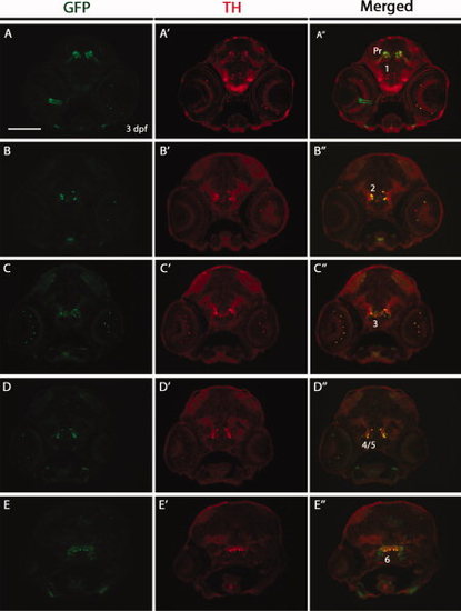
Double immunostaining for green fluorescent protein (GFP) and tyrosine hydroxylase (TH) on transverse cryosections of Tg(dat:EGFP) larvae at 3 days postfertilization (dpf). The 3 dpf Tg(dat:EGFP) embryos were transversely cryosectioned and stained with anti-GFP and anti-TH antibodies. GFP-positive cells and TH-positive cells are shown in green and red respectively. GFP expression in the optic nerve (A3) may relate to the previously reported DAT expression in astrocytes of the optic nerve (Holzschuh et al., 2001). Numbers (1–6) indicate different groups of DA neurons in the ventral diencephalon. Pr: pretectum. All sections are shown as dorsal to the top. Scale bar = 100 μm. |
| Genes: | |
|---|---|
| Antibody: | |
| Fish: | |
| Anatomical Terms: | |
| Stage: | Protruding-mouth |