|
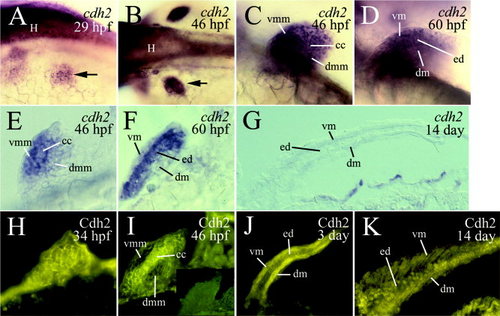
Expression of cadherin-2 mRNA (cdh2) and protein (Cdh2) in the embryonic zebrafish pectoral limb buds and larval fins. A–D are laterodorsal (A), dorsal (B), and lateral (C,D) views of the developing limb buds of 29 to 60 hours postfertilization (hpf) whole-mount embryos, labeled by in situ hybridization with cdh2 cRNA. Anterior is to the left. E and F are cross-sections (dorsal up) of the limb bud, whereas G is a horizontal section (anterior to the left) of the pectoral fin, showing cdh2 expression. H and I are cross-sections (dorsal up), whereas J and K are horizontal sections (anterior to the left) of the pectoral limb bud and fins, respectively, stained with Cdh2 antibody (immunofluorescent methods). The insert in I shows that preincubating the Cdh2 antibody with excess Cdh2-EC1-GST fusion protein (used to generate the antibody) blocked the labeling (Liu et al., 2001b). The arrows in A and B point to the left pectoral limb bud. Abbreviations are the same as in Figure 1.
|