
- Title
-
Cadherin-1, -2, and -11 expression and cadherin-2 function in the pectoral limb bud and fin of the developing zebrafish
- Authors
- Liu, Q., Kerstetter, A.E., Azodi, E., and Marrs, J.A.
- Source
- Full text @ Dev. Dyn.
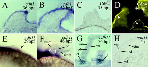
Expression of cadherin-1 message (cdh1; A,B), cadherin-4 immunoreactivity (Cdh4; C,D), and cadherin-11 message (cdh11; E–H) in the pectoral limb buds and fins in developing zebrafish. A–D are cross-sections of the limb buds (dorsal up). E and F are lateral views of the limb buds of whole-mount embryos (anterior to the left and dorsal up). G and H are horizontal sections of the pectoral fins (anterior to the left). The labeling of the epidermal tissue in D (arrowhead) is nonspecific, as demonstrated by experiments in which the epidermal labeling persisted while specific staining in the central nervous system was eliminated when the Cdh4 antibody had been preincubated with excess Cdh4 peptide used to generate the antibody (arrowhead in the insert; Liu et al., 2001a, b). The black arrow in E points to a few cdh11-positive cells. The white arrows in F indicate cdh11 staining in the proximal regions of the limb bud. The insert in G is a higher magnification of the basal region of the limb pectoral fin in G. cc, chondrogenic condensation; cl, cleithrum; dm, dorsal musculature; dmm, dorsal myogenic mesenchyme; ed, endoskeletal disc; vm, ventral musculature; vmm, ventral myogenic mesenchyme. |
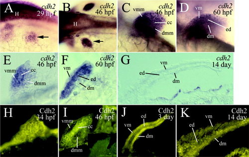
Expression of cadherin-2 mRNA (cdh2) and protein (Cdh2) in the embryonic zebrafish pectoral limb buds and larval fins. A–D are laterodorsal (A), dorsal (B), and lateral (C,D) views of the developing limb buds of 29 to 60 hours postfertilization (hpf) whole-mount embryos, labeled by in situ hybridization with cdh2 cRNA. Anterior is to the left. E and F are cross-sections (dorsal up) of the limb bud, whereas G is a horizontal section (anterior to the left) of the pectoral fin, showing cdh2 expression. H and I are cross-sections (dorsal up), whereas J and K are horizontal sections (anterior to the left) of the pectoral limb bud and fins, respectively, stained with Cdh2 antibody (immunofluorescent methods). The insert in I shows that preincubating the Cdh2 antibody with excess Cdh2-EC1-GST fusion protein (used to generate the antibody) blocked the labeling (Liu et al., 2001b). The arrows in A and B point to the left pectoral limb bud. Abbreviations are the same as in Figure 1. EXPRESSION / LABELING:
|
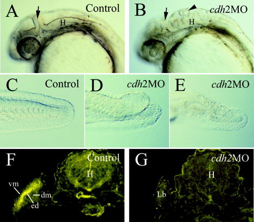
Embryos injected with cdh2 morpholino antisense oligonucleotides (cdh2MO) phenocopy the pac and the glass onion mutants. A–E: Views from 24 hours postfertilization (hpf) embryos with anterior to the left and dorsal up. B illustrates defects in the midbrain and hindbrain (H). The arrows in A and B point to the midbrain–hindbrain boundary. The arrowhead in B points to loose cell aggregates in the fourth ventricle. Compared with the tip of the tail in control embryos (C), cdh2MO embryos had split fin (D) or tail blister (E). F,G: Cross-sections (dorsal is up) of Cdh2 immunostaining of control morpholino oligonucleotide (Control) -injected (F) and cdh2MO-injected embryos (G), both at 50 hpf. dm, dorsal musculature; ed, endoskeletal disc; Lb, limb bud; vm, ventral musculature. |
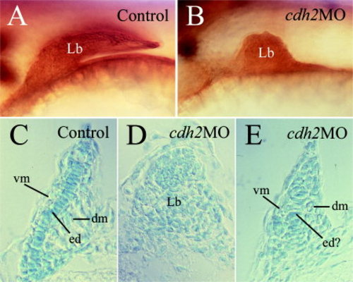
Effects of injection of cdh2 morpholino antisense oligonucleotides (cdh2MO) on the development of the pectoral limb buds. A,B: Lateral views of the pectoral limb bud of whole-mount embryos stained with an anti-Hu antibody (Sigma). This antibody labels differentiating neurons (Marusich et al., 1994). The staining in the limb buds, although nonspecific, makes their visualization easier. Dorsal is up and anterior is to the left. C–E: Cross-sections of limb buds (dorsal up) stained with Alcian blue showing anatomy of control (C) and cdh2MO-injected (D,E) embryos. ed?, possible endoskeleton; Lb, limb bud; Other abbreviations are the same as in Figure 1. PHENOTYPE:
|

Unillustrated author statements EXPRESSION / LABELING:
|