
Fig. 7
- ID
- ZDB-FIG-111011-9
- Publication
- Stevens et al., 2011 - Plasticity of photoreceptor-generating retinal progenitors revealed by prolonged retinoic acid exposure
- Other Figures
- All Figure Page
- Back to All Figure Page
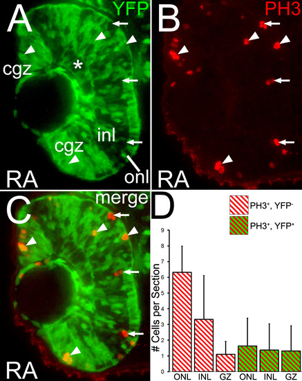
Proliferating cells in the retina can activate a reporter gene in response to RA. Embryos carrying the RARE-YFP transgene were treated with 0.3 μM RA at 36 hpf, and at 48 hpf were processed as 3 μm cryosections for indirect immunofluorescence with an anti-GFP antibody (green color; A) and a marker for mitotic cells, anti-phosphohistone H3 (red color; B); panel C shows the merged image. Dorsal is up. Asterisk indicates the ganglion cell layer (GCL). There are mitotic cells in the retina that are both negative (arrows) and positive (arrowheads) for YFP expression. (D) Numbers of singly-labeled (PH3+, YFP-) and doubly-labeled (PH3+, YFP+) mitotic cells were counted as a function of position in the retina in a sample of sections from RA-treated embryos. Cells counted in the developing ganglion cell layer were lumped together with those of the inner nuclear layer (INL). Single-labeled anti-PH3 cells are found largely in the outer nuclear layer (ONL) and INL. Few single-labeled anti-PH3 are seen in the GZ. Colabeled cells are found evenly distributed among these retinal regions. Bar = 50 μm. |