
Fig. 3
- ID
- ZDB-FIG-111011-6
- Publication
- Stevens et al., 2011 - Plasticity of photoreceptor-generating retinal progenitors revealed by prolonged retinoic acid exposure
- Other Figures
- All Figure Page
- Back to All Figure Page
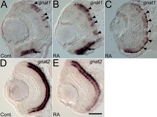
Expression of rod and cone transducin genes is altered by retinoic treatment beginning at the time of retinal neurogenesis. Embryos were treated with DMSO (Cont.; A, D), or 0.3 μM RA (B, C, E) at 36 hpf, and at 72 hpf were hybridized as 4 μm cryosections with probes corresponding to rod transducin (gnat1; A, B, C), or cone transducin (gnat2; D, E). Dorsal is up in all panels. In control embryos gnat1 is expressed in the ventral patch of rod photoreceptors (arrow in A) and in dispersed cells on the dorsal side of the retina (arrowheads in A). In some RA-treated embryos gnat1 expression is more intense in the ventral patch (arrow in B) and in all RA-treated embryos gnat1 expression is found in more cells in the dorsal and central retina (arrowheads in B and C). The section in panel C is positioned more peripherally in the retina compared to sections in other panels. Control embryos show an even distribution of gnat2 expression (D), which is reduced in intensity in RA-treated embryos (E). Bar = 50 μm. |