
Fig. 1
- ID
- ZDB-FIG-111011-4
- Publication
- Stevens et al., 2011 - Plasticity of photoreceptor-generating retinal progenitors revealed by prolonged retinoic acid exposure
- Other Figures
- All Figure Page
- Back to All Figure Page
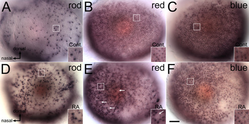
Opsin expression is altered by retinoic acid treatment beginning at the time of retinal neurogenesis. Embryos were treated with DMSO (Cont.; A, B, C), or 0.3 μM RA (D, E, F) at 36 hpf, and at 60 hpf were hybridized as whole mounts with probes corresponding to rod opsin (A, D), red cone opsin (B, E), or blue cone opsin (C, F); views are of whole embryonic eyes; dorsal is up and nasal to the left. In the control (DMSO-treated) embryos, rod photoreceptors are found predominantly in the ventral and dorsal regions (A), while red and blue cones (B, C) are evenly spread across the retina. In RA-treated embryos there is an apparent increase in rods, particularly in central regions of the retina (B), and a decrease in the appearance of red cones, leading to empty patches in the red cone mosaic (E, arrows). Boxed regions in each panel appear at higher magnification in the insets. Bar = 50 μm. |