
Fig. 6
- ID
- ZDB-FIG-111011-8
- Publication
- Stevens et al., 2011 - Plasticity of photoreceptor-generating retinal progenitors revealed by prolonged retinoic acid exposure
- Other Figures
- All Figure Page
- Back to All Figure Page
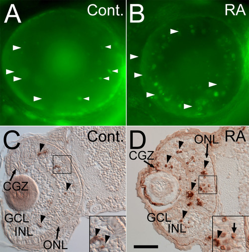
Retinoic acid treatment during retinal neurogenesis results in a significant increase in retinal cell death. Control embryos were treated with DMSO (A, C), or 0.3 μM RA (B, D) at 36 hpf, and either were stained live at 75 hpf with the cell death marker Acridine Orange and prepared as whole mounts for viewing (A, B), or were sectioned at 5 μm and processed for cell death detection with the TUNEL kit (C, D); dorsal is up in all panels. Control embryos showed very few dead/dying cells (arrowheads in A), and these were found predominantly in the inner nuclear layer (C), while those treated with RA (D) showed widespread cell death in the inner nuclear layer (INL), ganglion cell layer (GCL), and circumferential germinal zone (CGZ, D). However, little cell death was detected in the outer nuclear layer (ONL) (D). Bar = 50 μm for all panels. |