
Fig. 4
- ID
- ZDB-FIG-110915-12
- Publication
- Zygmunt et al., 2011 - Semaphorin-PlexinD1 Signaling Limits Angiogenic Potential via the VEGF Decoy Receptor sFlt1
- Other Figures
- All Figure Page
- Back to All Figure Page
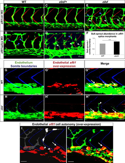
plxnD1 and sflt1 Interact Genetically, sflt1 Limits SeA Angiogenesis Cell Autonomously (A–I) Thirty-two hpf trunk vasculatures, green. (A–E) SBs, red. White arrowheads, ectopic SeA sprouts. Blue arrowheads, ectopic SeA branching. (A, C, E) Embryos treated with 20 ng of sflt1-ctrl MO: WT (A), obd/+ (C), obd (E). Embryos treated with 20 ng of sflt1-splice MO: WT (B), obd/+ (D). (F) 23 hpf SeA sprout abundance in WT (left, gray bar) and obd/+ (right, black bar) sflt1-splice morphants. n = 20 WT and n = 19 obd/+. Error bars represent SEM. ***p < 0.001. (G–I2) SBs, blue. GAL4FF/UAS-mediated endothelial-specific sflt1 overexpression, red. White arrows, missing SeA sprouts. (G2–H3) Endothelial sflt1 overexpression inhibits SeA sprouting. WT (G–G3). obd (H and H3), note lack of sflt1 overexpression (red) in remaining SeA sprout (white arrowhead). (I and I2) Mosaic vasculature with ECs from both obd donor and WT host. Endothelial-specific and mosaic sflt1 and DsRed coexpression restricted to the WT endothelium (red, I and I2). obd ECs express cytosolically targeted EGFP (gray in I; green in I2). WT ECs express nuclear-targeted EGFP (white in I; green in I2). obd and WT ECs without sflt1 overexpression (DsRed-) from SeA sprouts even next to sflt1 overexpressing WT ECs (DsRed+). WT ECs overexpressing sflt1 (DsRed+) fail to form SeA sprouts (white arrows, I and I2). (G–H3) n = 30 embryos with overexpression per genotype, all showing suppression of SeA sprouting. Anterior, left; dorsal, up. Scale bars represent 30 μm. |
| Fish: | |
|---|---|
| Knockdown Reagent: | |
| Observed In: | |
| Stage: | Prim-15 |
Reprinted from Developmental Cell, 21(2), Zygmunt, T., Gay, C.M., Blondelle, J., Singh, M.K., Flaherty, K.M., Means, P.C., Herwig, L., Krudewig, A., Belting, H.G., Affolter, M., Epstein, J.A., and Torres-Vazquez, J., Semaphorin-PlexinD1 Signaling Limits Angiogenic Potential via the VEGF Decoy Receptor sFlt1, 301-314, Copyright (2011) with permission from Elsevier. Full text @ Dev. Cell