
Fig. 1
- ID
- ZDB-FIG-110804-28
- Publication
- Lauter et al., 2011 - Two-color fluorescent in situ hybridization in the embryonic zebrafish brain using differential detection systems
- Other Figures
- All Figure Page
- Back to All Figure Page
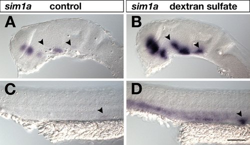
Effects of dextran sulfate in standard WISH. 24-hpf embryos were hybridized to a sim1a digoxigenin-labeled RNA probe, which was visualized by AP-BCIP/NBT chromogenic detection under identical conditions and staining times. Lateral views of embryonic brains (A,B) and trunks (C,D) are shown with anterior to the left. In (B,D) but not in (A,C) 5% dextran sulfate was included in the hybridization buffer. Addition of dextran sulfate resulted in increased signal sensitivity. Arrowheads in (A,B) indicate sim1a-positive neuronal clusters identified in dextran sulfate treated brains (B) that were hardly detected in untreated embryos (A). Arrowheads in (C,D) mark the pronephric primordium strongly visualized in dextrane sulfate treated embryos (D) but not in untreated specimens (C). Embryos were viewed on an Axioplan II microscope and images were recorded with an Axiocam digital camera. Scale bar is 100 μm. |